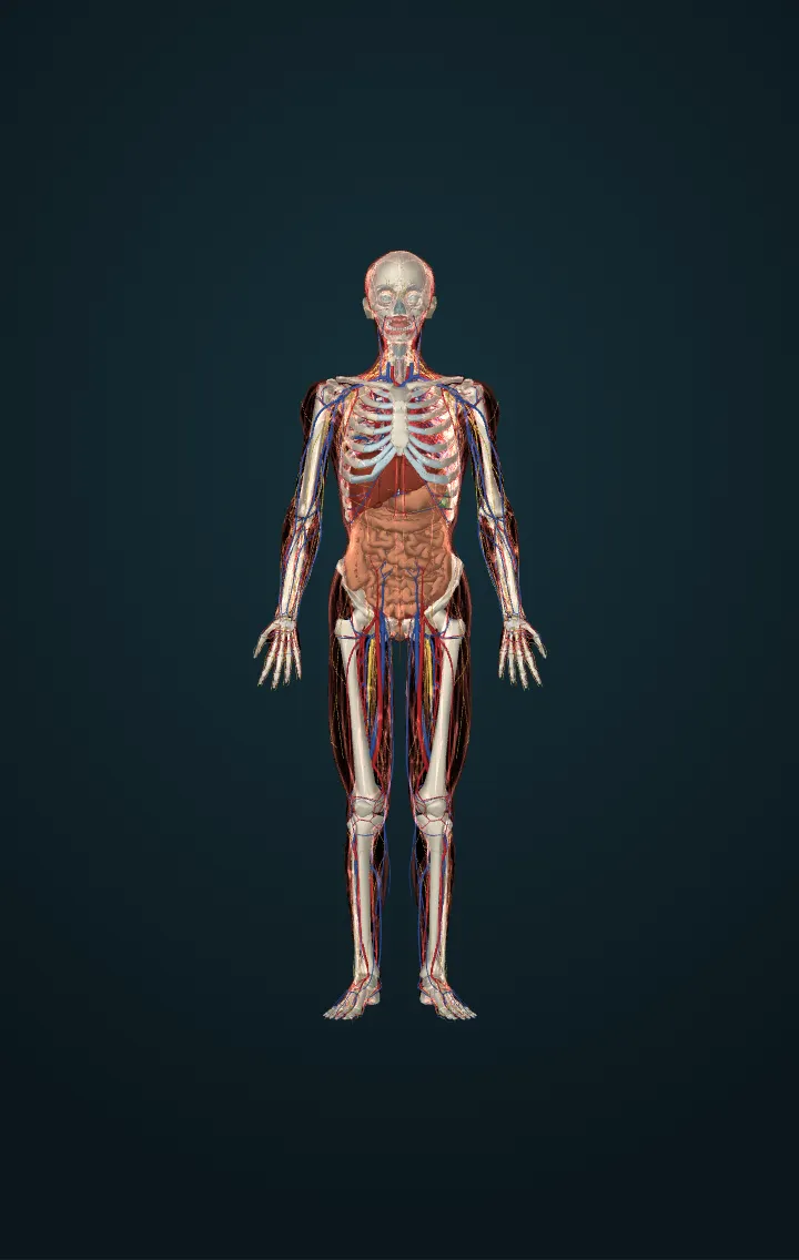
Female reproductive organs
The ovaries, the uterine tubes, the uterus, the vagina and the vulva: the organs that make eggs and the hormones estrogen and progesterone, bring egg and sperm together, and hold and nourish a pregnancy.
The female reproductive organs make eggs (oocytes), the female sex cells, and the hormones estrogen and progesterone; they bring an egg to where it can meet sperm, and they hold and nourish a growing embryo and fetus. Inside the pelvis are the two ovaries, the two uterine tubes (fallopian tubes), the uterus and the vagina; outside are the external genitalia, together called the vulva. The breasts, which make milk, are the system's accessory organs.
The ovaries, each about 2 to 3 cm long, the size of an almond, lie on either side of the uterus, tied to it by the proper ovarian ligament and to the wall of the pelvis by the suspensory ligament, which carries their blood vessels. A girl is born with one to two million immature eggs, each in a follicle. From puberty, about once every 28 days, one follicle ripens and releases its egg — ovulation — and what is left of it becomes the corpus luteum, which makes progesterone.
Each uterine tube runs from the uterus towards an ovary without touching it. Its funnel, the infundibulum, is fringed with finger-like fimbriae that sweep the released egg in. The tube's muscle and the beating cilia of its lining move the egg through the wide ampulla, where it is usually fertilised if sperm are there, and the narrow isthmus to the uterus.
The uterus, about 7 cm long and 5 cm wide when not pregnant, has a dome above the tubes' entries, the fundus; a body; and a narrow lower part, the cervix, which opens into the vagina. Its wall is mostly smooth muscle, the myometrium. Its lining, the endometrium, thickens every month to receive an embryo and is shed as the menstrual period when none implants. The vagina, a muscular canal about 10 cm long, leads from the cervix to the outside.
The vulva round the vaginal opening holds the labia, the openings of the urethra and the vagina, and the clitoris. The clitoris comes from the same embryonic tissue as the penis: its visible glans is only its tip, while its body and two crura, and the two vestibular bulbs beside the vaginal opening, lie under the skin and fill with blood when aroused. This page shows a woman's body, from the Human Reference Atlas's Visible Human female and an MRI model of a woman's pelvic floor, in place of the simulator's male body; the clitoris, the vestibular bulbs and glands, the labia minora and the ligaments are drawn by us where the anatomy texts place them.
Numbers
| Length of an ovary | about 2–3 cm | OpenStax, Anatomy and Physiology (1st ed., CC BY 4.0) — 27.2 Anatomy and Physiology of the Female Reproductive System |
|---|---|---|
| Size of the uterus, not pregnant | about 7 cm long and 5 cm wide | OpenStax, Anatomy and Physiology (1st ed., CC BY 4.0) — 27.2 Anatomy and Physiology of the Female Reproductive System |
| Length of the vagina | about 10 cm | OpenStax, Anatomy and Physiology (1st ed., CC BY 4.0) — 27.2 Anatomy and Physiology of the Female Reproductive System |
| Length of the menstrual cycle | 28 days on average, usually 21–32 | OpenStax, Anatomy and Physiology (1st ed., CC BY 4.0) — 27.2 Anatomy and Physiology of the Female Reproductive System |
| Immature eggs in the ovaries at birth | one to two million | OpenStax, Anatomy and Physiology (1st ed., CC BY 4.0) — 27.2 Anatomy and Physiology of the Female Reproductive System |
Its parts in the model
- Ampulla of left uterine tube
- Ampulla of right uterine tube
- Body of clitoris
- Body of uterus
- Cervix of uterus
- Endometrium
- External os of uterus
- Female urethra
- Fimbriae of left uterine tube
- Fimbriae of right uterine tube
- Fundus of uterus
- Glans of clitoris
- Infundibulum of left uterine tube
- Infundibulum of right uterine tube
- Internal os of uterus
- Isthmus of left uterine tube
- Isthmus of right uterine tube
- Isthmus of uterus
- Left bulb of vestibule
- Left crus of clitoris
- Left greater vestibular gland
- Left internal iliac vein
- Left internal pudendal vein
- Left labium minus
- Left ovary
- Left proper ovarian ligament
- Left round ligament of uterus
- Left suspensory ligament of ovary
- Left uterine artery
- Left uterine vein
- Right bulb of vestibule
- Right crus of clitoris
- Right greater vestibular gland
- Right internal iliac vein
- Right internal pudendal vein
- Right labium minus
- Right ovary
- Right proper ovarian ligament
- Right round ligament of uterus
- Right suspensory ligament of ovary
- Right uterine artery
- Right uterine vein
- Uterine horns
- Vagina
Sources
- OpenStax, Anatomy and Physiology (1st ed., CC BY 4.0) — 27.2 Anatomy and Physiology of the Female Reproductive System
- MedlinePlus — Female Reproductive System
- Human Reference Atlas, 3D reference organs of the Visible Human female (HuBMAP), CC BY 4.0 — adapted.
- Female Pelvic Floor Visualiser, © 2025 University of West Bohemia in Pilsen (Talimian, Čechová, Jansová, Hynčík), MIT licence — adapted.