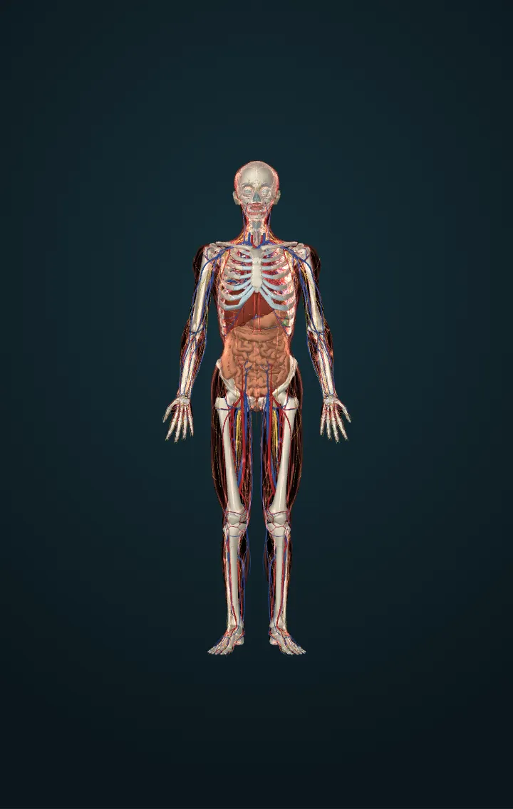
Mammary glands (breasts)
The glands in the breasts that make milk after a baby is born, and let it down to the nipple when the baby feeds.
The breasts are accessory organs of the female reproductive system: far from its other organs, their job is to feed a baby with milk, lactation. Each holds a mammary gland — a modified sweat gland — in fat and connective tissue, held up by suspensory ligaments; the fat, not the gland, decides a breast's size, and size does not decide how much milk it can make.
The gland is a set of lobes, each a cluster of lobules of tiny sacs, the alveoli, whose cells make milk. Ducts carry it to the nipple, where they open; round the nipple, the darker areola has small glands whose oily fluid protects it during feeding. The textbooks draw widened lactiferous sinuses behind the nipple; ultrasound of breastfeeding women found none, and fewer, smaller ducts that carry milk rather than store it.
In pregnancy, estrogen, progesterone, prolactin and other hormones make the ducts branch and the alveoli grow, while the placenta's hormones hold back milk-making until birth. In the first two or three days after birth the breasts give colostrum, thick and rich in protein and antibodies; mature milk follows from about the tenth day.
When the baby suckles, nerves in the areola set off a reflex: the pituitary releases prolactin, which makes milk for the next feed, and oxytocin, which makes the cells round the alveoli squeeze milk into the ducts — the let-down, within a minute of the baby starting. The more milk is taken, the more is made.
Numbers
| Areola's diameter | 25–100 mm | OpenStax, Anatomy and Physiology (1st ed., CC BY 4.0) — 27.2 Anatomy and Physiology of the Female Reproductive System |
|---|---|---|
| Feeds a day, a baby of 1–6 months fed only on breast milk | 11 on average (6–18) | Kent and others (2006), Volume and frequency of breastfeedings and fat content of breast milk throughout the day, Pediatrics 117: e387–e395 |
| Milk taken at a feed | about 76 g on average (0–240 g) | Kent and others (2006), Volume and frequency of breastfeedings and fat content of breast milk throughout the day, Pediatrics 117: e387–e395 |
| From suckling to the let-down | under a minute | OpenStax, Anatomy and Physiology (1st ed., CC BY 4.0) — 28.6 Lactation |
| Colostrum | the first 48–72 hours after birth | OpenStax, Anatomy and Physiology (1st ed., CC BY 4.0) — 28.6 Lactation |
Its parts in the model
- Areolar tubercles of left breast
- Areolar tubercles of right breast
- Lactiferous ducts of left breast
- Lactiferous ducts of right breast
- Lactiferous sinuses of left breast
- Lactiferous sinuses of right breast
- Left areola
- Left nipple
- Lobes of left mammary gland
- Lobes of right mammary gland
- Right areola
- Right nipple
- Suspensory ligaments of left breast
- Suspensory ligaments of right breast
Sources
- OpenStax, Anatomy and Physiology (1st ed., CC BY 4.0) — 27.2 Anatomy and Physiology of the Female Reproductive System
- Kent and others (2006), Volume and frequency of breastfeedings and fat content of breast milk throughout the day, Pediatrics 117: e387–e395
- OpenStax, Anatomy and Physiology (1st ed., CC BY 4.0) — 28.6 Lactation
- Ramsay, Kent, Hartmann and Hartmann (2005), Anatomy of the lactating human breast redefined with ultrasound imaging, Journal of Anatomy 206: 525–534
- Human Reference Atlas, 3D reference organs of the Visible Human female (HuBMAP), CC BY 4.0 — adapted.
- Female Pelvic Floor Visualiser, © 2025 University of West Bohemia in Pilsen (Talimian, Čechová, Jansová, Hynčík), MIT licence — adapted.